- Empty cart.
- Continue Shopping
Small Animal in Vivo Imaging System
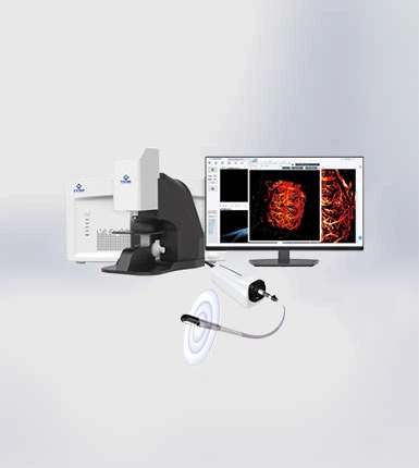
Small Animal In Vivo Imaging System GAni PA, GAni-Plus, GAni-OPO, GAni-OPO MAX Multi-modal (Photoacoustic, Ultrasonic) in vivo imaging Micron-level Resolution down to 3μm, Millimeter-level imaging depth up to 6mm 3D Merged Imaging.
Photoacoustic, ultrasound multimodal imaging
Photoacoustic imaging based on specific endogenous or exogenous light absorbing substances such as pigments, blood vessels, lipids, and nanoprobes
Ultrasound imaging based on acoustic impedance differences
Micron-level resolution, millimeter-level imaging depth
Photoacoustic microscopy breaks through the diffraction limit of traditional optical imaging,and the imaging depth is up to 6 mm.
At deeper imaging depths,high resolution at the optical level can still be maintained with an accuracy of 3 μm
3D image information is analyzed layer by layer
Through the real-time 2D tomographic data display overlay, the 3D structural images of the local tissue can be further obtained, and the 2D and 3D images can be further analyzed by using data processing software. |
Non-invasive, label-free imaging
Only a small amount of water (couplant) is applied to the imaging site to match the signal, and non-invasive imaging of the test site can be achieved without the injection of contrast agent. |
|||
Heating-anesthesia-integrated small animal fixation table
Integrated heating-anesthesia device specifically designed for better protection of model animals. |
Non-invasive, label-free imaging
Only a small amount of water (couplant) is applied to the imaging site to match the signal, and non-invasive imaging of the test site can be achieved without the injection of contrast agent. |
|||
PRODUCT PARAMETER
| Product name | Label-free multimodal in vivo imaging of small animals | |||
| Serial version | Standard Edition | Tunable wavelength version | ||
| Model | GAni Standard Edition | GAni-Plus Upgrade | GAni-OPO | GAni-OPO Ultimate |
| Imaging modality | Photoacoustic & Optical & Ultrasound Imaging | Dual-wavelength photoacoustic & ultrasound imaging | Photoacoustic & Ultrasound Imaging | Multi-wavelength photoacoustic & ultrasound imaging |
| Application direction | Brain, organs, tumors, blood vessels | Brain, organs, tumors, skin, blood vessels, pigments | Brain, organs, tumors, skin, molecular probes, blood vessels, pigments, NIR-I materials | Brain, organs, tumors, skin, molecular probes, blood vessels, pigments, lipids, NIR-I materials, NIR-II materials |
| Wavelength range | 532nm | 532nm& 1064nm | 532nm OPO(770-840nm) 1064nm | 532nm OPO(680-1190nm & 1150-2400nm) 1064nm |
| Imaging range | 3×3 mm, 1min | 3×3 mm, 1min | 3×3 mm, 1min | 3×3 mm, 1min |
| Imaging time | 20×20 mm, 20min | 20×20 mm, 20min | 20×20 mm, 20min | 20×20 mm, 20min |
| Lateral resolution | 3μm | 3μm | 3μm | 3μm |
| Axial resolution | 75μm | 75μm | 75μm | 75μm |
| Measurement depth | 3mm | 6 mm | 6 mm | 6 mm |
PRODUCT DESCRIPTION
GCell Multimodal small animal in vivo imaging system is a small animal in vivo imaging system that uses a variety of imaging technologies for comprehensive imaging, which can simultaneously detect and analyze the physiology, pathology, efficacy and other information of small animals. This technology can improve the accuracy and sensitivity of imaging, and provide more comprehensive and in-depth data support for biomedical research and drug development.
PRODUCT ADVANTAGE
GCell in vivo imaging system becoming increasingly popular due to their numerous advantages. Here are some of the most important benefits of this product:
1. Optical/photoacoustic/ ultrasound three-modal imaging
A three-modal in vivo small animal imaging system that integrates optical microscopy, photoacoustic imaging of endogenous light-absorbing substances such as pigments and blood vessels, and ultrasound imaging of acoustic impedance differences.
2. Micron-level resolution, millimeter-level imaging depth
Micron, high-resolution imaging of tissue structures within 3 mm can still be performed without the need for contrast media, and the position of the focus can be adjusted according to the real-time display of the software.
3. Three-dimensional image information is analyzed layer by layer
Through the real-time 2D tomographic data display overlay, the 3D structural images of the local tissue can be further obtained, and the 2D and 3D images can be further analyzed by using data processing software.
4. Non-invasive, label-free imaging
Only a small amount of water (couplant) is applied to the imaging site to match the signal, and non-invasive imaging of the test site can be achieved without the injection of contrast agent.
5. Heating-anesthesia-integrated small animal fixation table
Integrated heating-anesthesia device specifically designed for better protection of model animals.
6. Imaging systems with customized light sources
According to the different needs of customers, customize the corresponding single-wavelength, multi-wavelength, tunable wavelength light source imaging system.
PRODUCT APPLICATION
GCell in vivo imaging system are widely used in below area
1. Monitoring of tumor growth process
The monitoring of the growth of tumor trophic blood vessels in the ears of mice, the monitoring of the growth of tumor trophic blood vessels, and the relationship between the curvature, density and depth of tumor trophic blood vessels and the tumor growth time were verified.
References
[1]. F. Yang, et al..J. Biophotonics, e202000022.2020.DOI:10.1002/-jbio.20000022
[2]. Z. Wang, Nanophotonics,10(12), 3359-3368, 2021.DOI:10.1515/nanoph-2021-0198.
2. Monitoring of the treatment process of tumors
The monitoring of the ablation of the nourishing vessels during the photodynamic (PDT) treatment of back tumors in mice was realized, and the relationship between the curvature, density and depth of the tumor trophic vessels and the duration of PDT treatment was revealed.
References
F. Yang, et al.,J. Biophotonics, e202000022.2020, DOI:10.1002/-jbio.20000022.
3. Functional imaging of the brain in small animals
The dynamic monitoring of “ischemia-reperfusion” of the vascular network deep in the mouse brain was realized, and the broad application prospect of this instrument in the basic research of cerebrovascular diseases was demonstrated.
References
F.Yang. et al.. J. Biophotonics, e202000022.2020.DOI:10.1002/- jbio.20000022
4. Assess the extent of blood supply to the lesions
The evaluation of the degree of blood supply to the back of mice and the total retreat of mice was realized, which broke through the bottleneck of imaging technology to evaluate the degree of blood supply to damaged tissues and improved the possibility of rapid surgical intervention.
References
D.Zhang.et al., Quant Imaging Med Surg, 11(10).4365-4374.2021.DOI:10.21037/qims-21-135.
5. Imaging of iris and sclera in live animals
It can realize the imaging of the iris and scleral vascular network of the eyes of live small animals (such as mice) and large animals (such as rabbits).
6. Nanoprobes and molecular imaging studies
Tumor-specific photoacoustic imaging at special wavelengths (custom version)
The photoacoustic multi-modal small animal imager can be customized, and the specific nanoprobe can be used to improve the amplitude of the photoacoustic imaging signal of the tumor area for special wavelengths, so as to achieve large-depth and high-sensitivity tumor-specific photoacoustic imaging.
References
[1]. D.Cui, et al.. Nano Letters, 21(16).6914-6922.2021, DOI:10.1021/acs. nanolett.1c02078[2]. J.Zheng. et al., J. Am. Chem. Soe,141(49),19226-19230.2019.DOI: 10.1021/jacs.9b10353.
7. Breast tumor specimen marker imaging
T.Wong.et_x0001_al.. _x0001_Sci.Adv.,3_x0001_(5)._x0001_e1602168.2017.D01:_x0001_10.1126/sciadv.1602168.
Labeled imaging of liver micrometastases in early-stage neoma
Q.Yu,et_x0001_al.,J_x0001_Nucl_x0001_Med. 61(7),10791085,2020.00I:_x0001_10.2967/inumed.119.23315
8. Ambulatory monitoring of structural and functional changes in the early stages of abscient stroke
J.Lv.et_x0001_al.,_x0001_Theranostics,10(2).816-828.2020.DOI:10.7150/thno.38554.
Multimodal imaging observations of the living eye before and after suture injury
J.Park.B.Park.et_x0001_al.,_x0001_PNAS.118(11)._x0001_e1920879118.2021,_x0001_DO1:10.1073/pnas.1920879118.
Imaging of the retina in live animals, choroid, iris, sclera
C.Tian,_x0001_et_x0001_al.,_x0001_0ptics_x0001_Express,25(14)._x0001_15947-15955,2017.DOI:10.1364/0E.25.015947.
Z.Hosseinace,_x0001_et_x0001_al.,_x0001_Optics_x0001_Letters,45(22).6254-6257,2020.DOI:10.1364/0L.410171.
Labeled imaging of cells in the liver
D. Deng.et_x0001_al.,Nanophotonics,2021,DOI:/10.1515/nanoph-2021-0281.
9. Quantitative assessment of pigment distribution
The photoacoustic multimodal imaging system can quantitatively assess skin pigmentation and assist in clinical diagnosis
References
H.Ma. et al., Appl, Phys, Lett.. 113,083704,2018.DOI:10.1063/1.5041769.
10. Microvascular quantitative assessment
The photoacoustic multimodal imaging system can quantitatively monitor the effect of bright erythema before and after treatment, and give the most intuitive feedback on pathological parameters
Reference
H. Ma. et al.. Bio. Exp.12(10).6300-6316.2021.DOI:10.1364/B0E.439625.
Two-dimensional assessment Three-dimensional quantification Pre- and post-treatment evaluation


