- Empty cart.
- Continue Shopping

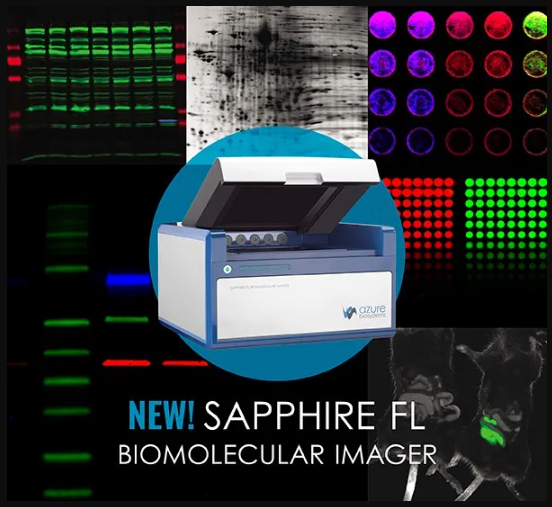
Easy Screening of Autophagy-Associated Acidic Compartments with the Sapphire Biomolecular Imager
- Posted on
LabmallX
- Categories: Bulletin & Updates,Library


What is autophagy? Autography is the homeostatic cellular mechanism by which unneeded cellular components are degraded by lysosomes. Disruption of the autophagy pathway contributes to the development of a variety of human diseases. Mechanistically, the autophagic pathway includes the formation and maturation of a series of different acidic vesicles that eventually converge in the lysosome for cargo degradation.
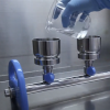
In this new application note, screening for autophagy activity in cells stained with Acridine orange (AO) acidotropic fluorescent dye is described and detected with a Sapphire Biomolecular Imager. AO is a cell-permeable fluorescent dye that accumulates in acidic compartments, such as lysosomes and autophagic and endosomal vesicles, allowing their detection in live cells.
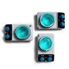
Green to red fluorescence intensity ratio for cells submitted up to 1, 2, or 3 hours of starvation with 1 or 2 µg/ml in µg/ml AO (left). Image of multi-well plate with cells subjected to 1, 2, or 3 hours of starvation (STV1, STV2, STV3 respectively), stained with 1 or 2 µg/ml AO, and incubated with or without Chloroquine (right).
This app note was made in collaboration with the Grasso Lab from the University of Buenos Aires in Argentina.
The Sapphire FL is the ultimate biomolecular imager for flexibility. With customizable and user-changeable laser and filter modules, it effortlessly adapts to your changing needs and advancing research.